Summary: The FDA has cleared a dental AI platform for radiograph analysis, emphasizing the importance of its integration into clinical workflows. Dental practices should ensure the AI system supports imaging, CBCT review, voice charting, and claim documentation to reduce bottlenecks. FDA clearance confirms the product's intended use, but practices must verify the AI's applicability to their specific imaging needs and patient groups.
A 2025 umbrella review hosted by PMC reported pooled AI sensitivity of 0.85 and specificity of 0.90 for caries detection. That matters because a rushed bitewing on a busy hygiene column is no longer just a human attention problem. The same radiograph can slow the doctor, weaken the patient explanation, and leave the insurance narrative short of what the payer needs. An FDA-cleared dental AI platform for radiograph analysis should be judged by what it changes after the image appears on screen. The defensible position is simple. Practices should not buy dental AI as a highlight-box feature. A clinical review system earns its place when imaging, CBCT review, voice charting, and claim documentation cut the same bottleneck instead of creating separate AI chores.
A dental owner choosing automation is really choosing where clinical judgment gets support, where staff time stays protected, and where the record becomes easier to defend. The sections below treat FDA clearance as the starting gate, not the finish line. The platform still has to work inside a real appointment.
What FDA clearance actually tells a dental clinic
FDA clearance answers a narrow but important question. The product has been reviewed for a specific intended use, and the practice should read that intended use before assuming the software covers every radiograph, patient group, or finding. A cleared caries aid, a panoramic pathology detector, and a CBCT periapical radiolucency tool are not interchangeable. The first procurement task is matching clearance language to the images the practice already takes.
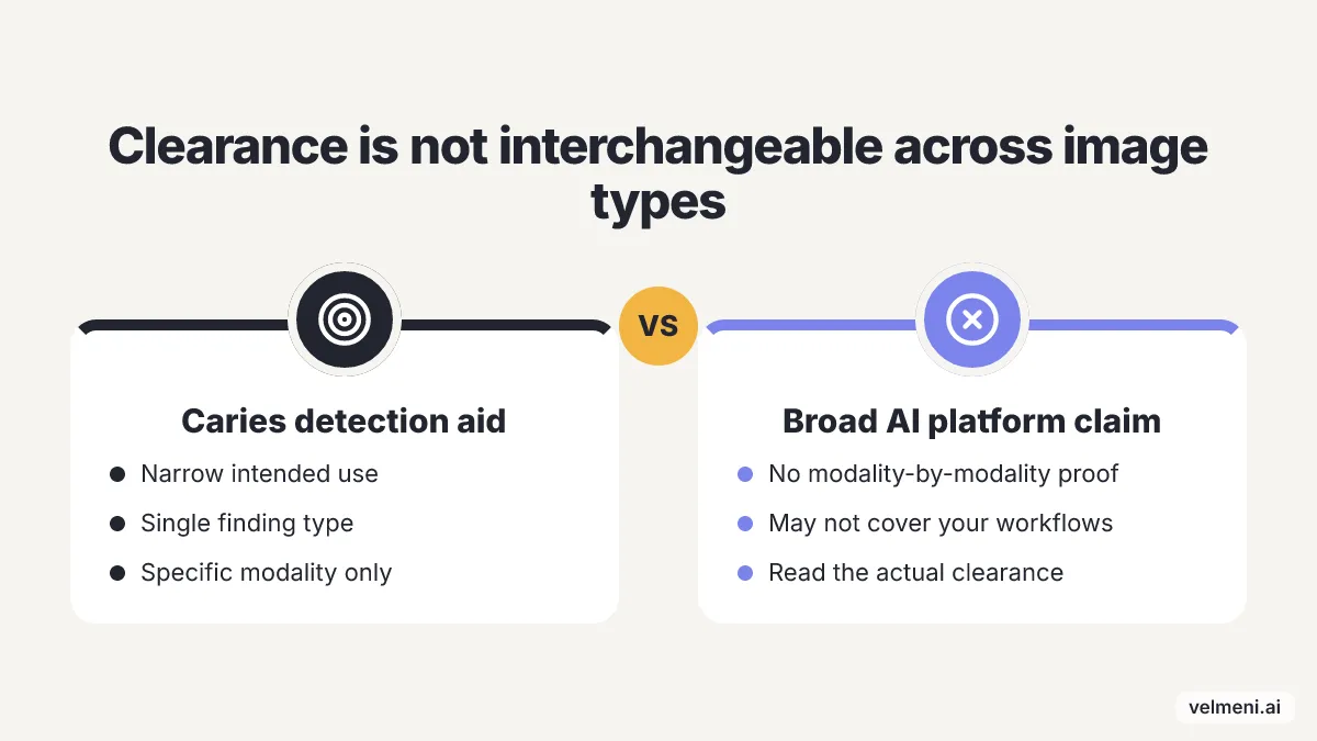
Radiograph automation has moved quickly from single-surface detection toward broader review. The April 2026 FDA listing for Dentsply Sirona shows DS Core CBCT Anatomy cleared as automated radiological image processing software, while the January 2026 Diagnocat clearance letter describes concurrent-read detection software to aid detection of periapical radiolucency on CBCT images. Those clearances show why a practice should ask for modality-by-modality coverage instead of accepting a broad AI label.
Recent clearances a practice may hear about
| Product or clearance | Month and year | Image or finding named in the source | Practical question for the practice |
|---|---|---|---|
| Dentsply Sirona DS Core CBCT Anatomy | April 2026 | The FDA database lists automated radiological image processing for the product. | Does the tool support the CBCT review your clinicians actually perform, or only a defined processing task? |
| Diagnocat K252934 | January 2026 | The FDA clearance letter describes periapical radiolucency on CBCT as the aided detection use. | Will the AI run as a concurrent read before the doctor signs the radiographic interpretation? |
| Dentsply Sirona DS Core Detect | January 2026 | Elegantlab reported clearance for incidental CBCT findings involving teeth with periapical radiolucency in adults 22 and older. | Are incidental findings routed into the chart and patient conversation, or left as a separate screen? |
| Pearl Second Opinion panoramic expansion | December 2025 | Pearl announced FDA clearance for panoramic radiograph detection of dental pathologies. | Does the practice need panoramic support because full-mouth explanations are part of the consult? |
Clearance should be read like a clinical scope document. A practice that buys software for panoramic imaging should not assume the same platform has the same clearance for bitewing caries, periapical images, or CBCT volumes unless the vendor can show the exact intended use. That habit protects the dentist from two common procurement mistakes. One is buying a narrow tool for a broad workflow. The other is rejecting a useful tool because it does not claim more than its clearance allows.
Where should AI enter the clinical review workflow?
The best place to automate is before the doctor starts falling behind, because delays in image review tend to leak into the rest of the appointment. A practical radiograph workflow puts AI after image capture, before the dentist gives the final read, and before the finding turns into a patient explanation or claim narrative. Image capture should start the first review layer, without sending an assistant off to upload files into a second system. The order that actually works is capture, AI analysis, clinician confirmation, patient explanation, chart entry, and claim support. If a platform forces staff to copy findings from one window into another, it saves less time than it promises, because the practice still pays for the handoff work.
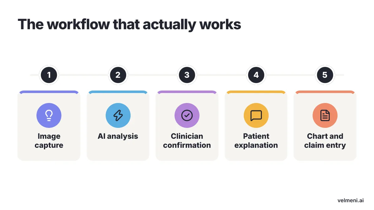
The clinician still owns the diagnosis, and the software should make that ownership easier to document. It should show what it flagged, let the dentist accept or reject the finding, and keep enough context for the record. A chairside tool that cannot support the chart note or claim attachment leaves the practice with a visual prompt and no finished workflow. VELMENI is positioned as the industry's only unified AI platform combining 2D/3D imaging, claims and voice into one FDA-cleared dental system. In practical terms, VELMENI provides AI X-ray & imaging analysis (2D: panoramic, bitewing, periapical), 3D/CBCT AI analysis and verification, and VELMENI Voice (voice-driven perio charting), which puts the review, charting and documentation jobs inside one platform rather than treating them as separate purchases.
Voice-driven perio charting belongs in the same discussion as radiograph AI, because clinical review automation falls apart when the doctor still has to stop for manual data entry. A perio exam charted by voice cuts keyboard dependence during the appointment, while imaging AI keeps the radiographic review visible for the doctor and patient. The next procurement question is whether the platform can handle everyday 2D review and higher-stakes CBCT work without splitting the team across tools.
Why 2D and CBCT review should not be bought separately
Separate 2D and CBCT tools create hidden training costs when different interfaces, evidence displays, and reporting habits compete inside the same practice. General dentists, oral surgeons, and implant teams may read different studies, but the patient record needs one coherent story from screening image to treatment plan.
Diagnostics published a 2024 systematic review finding AI caries-detection sensitivity across studies ranged from 0.360 to 0.940, while specificity ranged from 0.700 to 0.983 across clinical imaging modalities. That spread is why a practice should test AI on its own image mix instead of asking only whether the product has AI. The question is how the tool performs on the radiographs, sensors, positioning habits, and case types already in the practice. CBCT review changes the stakes because 3D volumes are packed with information and slow to review consistently. The January 2026 Diagnocat clearance for CBCT periapical radiolucency detection and the April 2026 DS Core CBCT Anatomy listing show that automated 3D review is becoming a defined category, not a speculative add-on. A clinic that already takes CBCT for implants, endodontics, or oral surgery should test 3D/CBCT AI analysis and verification alongside its 2D workflow, because the review burden does not stop at bitewings.
A single-chair practice that takes only bitewings, does not place implants, and does not struggle with image-supported claims should not buy a unified imaging, voice, and claims platform first. For that practice, a focused caries-assist tool plus a written radiograph review protocol is the cleaner test, because the narrower workflow has fewer handoffs to fix. The case for one platform gets stronger when the same team is managing 2D screening, CBCT review, perio charting, patient education, and claim documentation.
A platform decision should therefore include a live charting and reporting test, not just a detection demo. Ask the vendor to process a typical bitewing series, a panoramic radiograph, and a CBCT case, then have the doctor explain the findings to a staff member acting as the patient. The weak point shows up fast when the team tries to move from AI flag to accepted treatment plan to payer-ready record.
How does clinical review automation change the patient conversation?
Patient understanding changes when the finding is visible, labeled and connected to the proposed treatment at the same appointment. Radiograph AI is not a substitute for the dentist's explanation, but it gives the explanation a shared visual reference that reduces the gap between what the doctor sees and what the patient understands.
Immersive VR 3D/CBCT visualization for patient education is most useful when the patient needs to understand spatial relationships, not only a single highlighted area. Implant planning, impacted teeth, airway discussions and complex pathology reviews depend on orientation in a volume, and a flat screenshot can make those cases harder to explain. A practice using CBCT should therefore ask whether the patient education tool can show the volume in a way that supports consent and treatment discussion.
AI-assisted insurance claims support belongs in the same workflow because the patient conversation and the payer record should be built from the same clinical evidence. A radiographic finding that is accepted by the dentist can feed the narrative, images and documentation package more reliably when the system is designed for claims support instead of being a diagnostic viewer alone. The operational benefit is not a promise that every payer will approve every claim, but a cleaner record that reduces avoidable back-and-forth caused by missing or inconsistent documentation.
Cone beam reporting is not just a document at the end of the process. The report is where findings, clinical review and referral communication become durable enough for future treatment planning, so the platform should make reporting part of the workflow rather than an after-hours task.
Patient education, claims and reporting are therefore not soft extras around radiograph AI. They are the places where missed handoffs show up as declined treatment, staff rework or incomplete records. A practice should judge the system by whether the finding can move from image to chairside explanation to documentation without being retyped by three different people.
What should a practice test before signing?
Demo days go better when you bring the messiest cases you actually see. If chairside X-ray reads are slowing the day down, you will probably end up comparing two setups: a narrow tool that reads one image type, or broader software that ties bitewings and CBCT data straight into charts and insurance packets. The best trial uses your normal patient scans and makes everyone finish a full appointment cycle while the sales team sits there and watches.
Bring the whole crew into the room for this step. Put the dentist, the hygienist, the dental assistant, your treatment coordinator, and whoever handles billing right at the screen. The dentist should be judging how quickly they can reject a bad read. The hygienist needs to see whether pocket depth entry drags down the schedule. Your billing person has to check whether the clinical notes match the rules your payers actually demand. A polished screen does nothing if someone has to rebuild the record by hand after five o'clock.
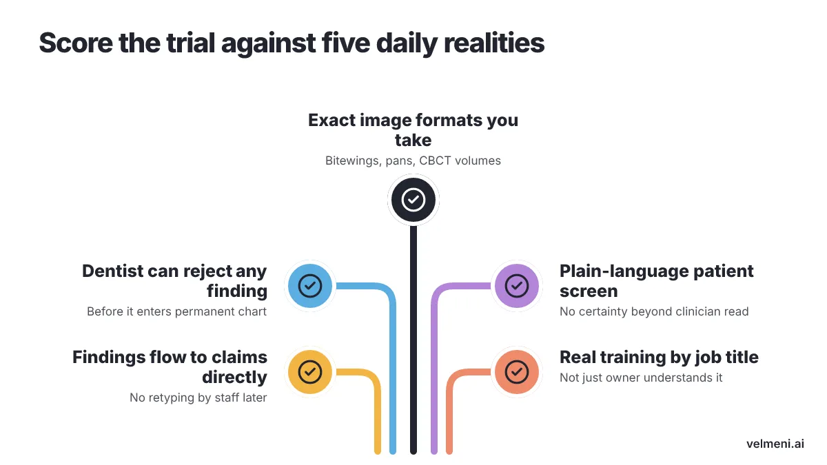
Score the trial against the things your team handles every day:
- The software has to read the exact image formats you take each week, whether that means bitewings, standard periapicals, pans, or three-dimensional CBCT volumes.
- Dentists need the ability to change, approve, or toss out any suggested finding before it lands in the permanent chart.
- The patient-facing screen should explain the problem in plain language without sounding too certain or getting ahead of the clinician.
- Accepted evidence needs to move straight into claim documentation, so staff members are not writing the same finding twice.
- The company should give a clear training schedule for every job title in the building, because software only the owner understands does not fix the daily schedule.
Dental offices inside the United States do not have to stop at a regional X-ray AI provider, as long as the platform connects with existing sensors, fits the charting steps already in place, and comes with real staff training. You want to see whether that vendor will let you run ordinary patient scans through the engine before you sign a contract.
Run a small test drive in your own operatory instead of making an open-ended promise to buy new technology. Gather recent scans, name the staff members who will check the software suggestions, write down the path an accepted finding takes into the chart, and compare the claim paperwork with the way you file today. If the system trips during that run, walk away, no matter how sharp the canned demo pictures looked.
Conclusion
Lay out the path from start to finish. You want to see where an X-ray gets taken, when the dentist reads it, how you present it across the chair, and how the billing office packages the claim. A solid trial depends on real patient files, your actual staff, and a software company willing to let you follow an image all the way to the final insurance packet. Set up a meeting with a vendor once you decide to look past simple radiograph overlays. Hand them a bitewing run, a panoramic scan, a tricky CBCT volume, and one denied claim that took weeks of phone calls to fix. Choosing the software is where software advice stops. Diagnosis, treatment decisions, and billing accuracy stay with the licensed staff in the office.
Where to go next
Related reading
- How to Reduce Rejected Dental Claims Using AI Evidence
- Voice Dental Charting: Safer Multilingual Notes
Sources
- PMC — "Examining the diagnostic accuracy of artificial intelligence for detecting dental caries across a range of imaging modalities: An umbrella review with meta-analysis"
- FDA: 510(k) clearance for Dentsply Sirona's DS Core CBCT Anatomy
- FDA: 510(k) clearance letter for Diagnocat (K252934)
- Elegantlab (citing FDA 510(k)): Dentsply Sirona's DS Core Detect cleared for incidental CBCT findings
- Pearl (hellopearl.com): Second Opinion AI expanded with FDA clearance for panoramic X-rays
